- Healthcare Professionals
- Oncology
- Diagnostics
- Tests and scans
Pathology tests
Pathology
Within our network of state-of-the-art centres, we offer a comprehensive pathology service covering many different specialities, including breast, prostate and gynaecology. We use the latest proven screening and diagnostic techniques, conducted by experienced clinicians.
The pathology tests available at GenesisCare are listed below and you can read more on our patient site.
What we offer
- Biopsy (Fine needle aspiration), core biopsy, image-guided biopsy, TRUS biopsy, transperineal biopsy)
- Blood testing
- Colposcopy
- Cystoscopy
- Smear test
- Urine cytology tests
- Urodynamic tests
Radiology tests
Radiology
Our expert nuclear medicine physicians, radiographers and radiologists use the latest-generation technology to perform and interpret the scans. As well as established methods of scanning, we also use innovative solutions to identify disease and plan your treatments, such as PSMA PET/CT for prostate cancer and fMRI to examine the brain.
The radiology services available at GenesisCare are listed below and you can read more on our patient site.
What we offer
- CT
- Digital X-ray
- Digital mammography
- Fluoroscopy
- MRI (Including fMRI and mpMRI)
- PET/CT (Including PSMA PET/CT and SPECT/CT)
- Ultrasound (Including transrectal ultrasound and transvaginal ultrasound)
Genetic and genomic testing
Genetic and genomic testing
We’re pioneering the use of genomic profiling in the diagnostic pathway for all solid tumours, leukaemias, lymphomas or sarcomas.
We provide genetic testing to patients undergoing cancer treatment at GenesisCare, if their clinician feels it's appropriate. We do not offer genetic testing outside of a treatment pathway.
MR Imaging in neuro-oncology
Multi-modal MR imaging guides cognitive and neurological outcomes for patients with primary brain tumours and brain metastases.
Functional MRI
Functional MRI (fMRI) is a non-invasive imaging technique that is able to indirectly measure brain activity using fluctuations in brain energy demands related to neural signalling.1
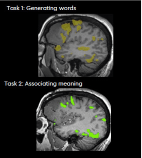
We use fMRI to locate eloquent brain tissue related to speech, vision, movement, etc., as well as guide neurosurgical planning and tailor surgery for intrinsic brain tumours to avoid neurological deficit and improve patient outcomes. With the increasing neuro-oncological benefits of surgery for brain tumours, fMRI offers potential further applications to measure brain recovery processes to optimally stage and monitor treatments.2
Generally, fMRI shows high agreement with clinical gold standard tests, such as the amytal test and intraoperative direct brain stimulation, but importantly offers the advantage of being safe and non-invasive.
Diffusion tensor imaging
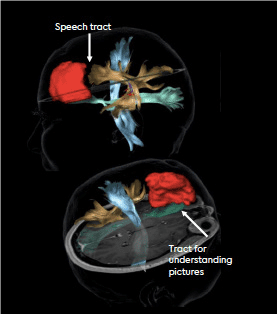
In addition, patients are offered a complementary MRI technique called diffusion tensor imaging (DTI). This imaging scan provides assessment of tissue microstructure and we use tractography to provide a 3D visualisation of neural tracts and the brain’s connectivity. This provides additional information about the location of important pathways for speech, movement, vision and other behaviours important for patient quality of life, whereas conventional MRI techniques provide only anatomical information.
These innovations that preserve quality of life are crucial to our approach to neuro-oncology which is to treat the whole patient, and not just the tumour. We are the first private service to offer this to our patients in the UK.
DTI determines proximity of language tracts

fMRI determines proximity of language activity

Refer to us
Oxford
If you would like further information about our neuro-oncology service, please contact rem@genesiscare.co.uk
References:
- Glover et al., 2011; Neurosurg Clin N Am; 22(2):133‑139.
- Brown et al., 2016; JAMA; 316 (4):401-409.
PET/CT scans
Our advanced imaging techniques and treatments help us to provide the best possible care and personalise your patient’s treatment to meet their exact needs. At GenesisCare, we aim to provide PET/CT scans within 48 hours of referral and send the results within 24-48 hours of completing the scan.
At GenesisCare, we offer a range of radioisotopes used with PET/CT scans at our Oxford and Windsor centres.
The key types of PET/CT scans we offer are:
- 68Gallium PSMA PET/CT
- 18F PSMA PET/CT
- 18F FDG PET/CT
- 68Gallium Dotatate PET/CT
At GenesisCare, we aim to provide PET/CT scans within 48 hours of referral and send the results within 24-48 hours of completing the scan.
On-site radiopharmacy
At GenesisCare, we are proud to have our own onsite radiopharmacy at our Windsor centre. A significant obstacle when using PET/CT scans is obtaining the radiopharmaceuticals used in some of the scans. This is made especially difficult due to the short half-lives of some of the radiotracers.
Having our own radiopharmacy eliminates this difficulty as we can produce our own 68Gallium PSMA and 68Gallium Dotatate labelled radioisotopes onsite. This means we can provide our patients with fast access to the diagnostics scans essential to their treatment, without delay.
We provide fast access to our comprehensive cancer treatment pathways if needed, with treatment often starting within days of diagnosis.
Range of radiotracers and imaging techniques
Our range of advanced and innovative imaging techniques provide highly precise and detailed scans that help guide our diagnosis and treatment pathways, so we can achieve the best possible outcome for your patient.
Outpatient oncology specialists
Our purpose-built centres at Oxford and Windsor have been awarded the Macmillan Quality Environment Mark (MQEM) and offer some of the UK’s most advanced cancer therapies. Our highly experienced multidisciplinary teams will care for your patient from their very first scan through to treatment and aftercare.
We offer a complete care plan that is designed to support your patient physically and emotionally, including exercise medicine and wellbeing therapies in partnership with Penny Brohn UK.
Refer for PET/CT scans
To enquire about PET/CT scans or to refer a patient, please call us or send an email.
Types of PET/CT Scans
By using different radiotracers, we can diagnose and monitor a range of cancers.
68Gallium PSMA PET/CT scans are the gold standard imaging scan for prostate cancer. 68Gallium is combined with a unique carrier molecule to create the radiotracer. The carrier molecule is attracted to prostate-specific membrane antigen (PSMA). PSMA is a protein that is found on the surface of prostate cancer cells in high concentrations. This includes cancer cells that may have spread to other parts of the patient’s body. 68Gallium PSMA is an effective radiotracer used in diagnosing prostate cancer due to its increased specificity with low PSA (prostate-specific antigen) levels and sensitivity to tumour, lymph and bone metastases.
A 68Gallium PSMA PET/CT scan is also superior to traditional bone imaging and can distinguish between bone damage caused by cancer and other non-cancer causes. We can closely and accurately monitor how well prostate radiotherapy or hormone therapy cancer treatment is progressing for a patient.
Theranostics – 177Lutetium PSMA therapy
We use 68Gallium PSMA PET/CT scans as the first step in our ‘seek and destroy’ Theranostics treatment for metastatic castration-resistant prostate cancer. The 68Gallium PSMA scan will show us if your patient is suitable for therapy using 177Lutetium PSMA and will be used to assess how well it’s working. If suitable, we will use the radioactive substance 177Lutetium combined with PSMA to target the prostate cancer cells. Its unique ability to seek out the exact location of your patient’s cancer and any areas it may have spread to, means that treatment can be focused directly on these areas.
18F PSMA PET/CT
We also use 18F PSMA PET/CT at our Bristol centre through a fortnightly mobile unit. This works the same way that 68Gallium PSMA PET/CT scans do, but the radiotracer used is 18Fluorine. This radiotracer is easy to transport, which means we can offer these scans to more patients at a mobile unit.
You can refer for 68Gallium Dotatate PET/CT scans which are used to diagnose and assess primary and metastatic well-differentiated neuroendocrine tumours (NET) particularly in symptomatic patients with negative anatomical findings. As NET tumours overexpress somatostatin receptors, Dotatate, which is an analogue of somatostatin, is a carrier molecule for the 68Gallium and provides a highly sensitive method for identifying NETs throughout the body. This scan also plays a key role in identifying if a patient may benefit from a Theranostics pathway by highlighting disease areas and suitability for 177Lutetium Octreotate treatment.
18F FDG PET/CT scans are also available at our Windsor and Oxford centres and are used to detect and access the metabolic activity of certain tumours including lung cancer, colorectal cancer, lymphoma, melanoma, breast cancer, ovarian cancer, brain cancer and multiple myeloma. FDG (fluorodeoxyglucose) is similar to naturally occurring glucose which all the cells of the body use for energy. As cancerous cells use more glucose than normal cells, this enables the 18F FDG PET/CT scan to demonstrate areas of increased uptake, making it an ideal imaging tool at diagnosis and for follow-up of treatment.
Work with us
If you’re a consultant and would like to work more closely with us and provide diagnostics or treatment, you can apply for practising privileges at one of our centres.
Make a referral
Referring a patient to GenesisCare is easy. Even if you don't have practising privileges, you can still refer directly for diagnostics or refer to one of our surgeons or oncologists for treatment. Contact us today to refer a patient for fast access to advanced diagnostics.
SPECT-CT scan
Radionuclide imaging including SPECT-CT
Our advanced imaging techniques and treatments help us to provide the best possible care and personalise your patient's treatment to meet their exact needs. At GenesisCare, we aim to provide radionuclide scans, including SPECT-CT scans, within five working days of referral and send the results within 24-48 hours of completing the scan.
Radionuclide imaging
Radionuclide imaging is highly useful in clinical oncology, collecting precise information to help our expert consultants guide diagnosis and treatment pathways.
The key types of scans we offer, which are available with or without a SPECT-CT, are:
- 99mTc HDP bone scan
- 123I whole body scan
- 99mTc DMSA renal scan
The range of these scans with optional SPECT-CT enables us to detect and monitor a wide variety of conditions, including cancers.
These scans are available at our Windsor centre.
Refer for radionuclide imaging
To refer a patient or enquire, please send an email to the below.
Why refer?
Fast turnaround time
We know how important time is when it comes to cancer diagnosis and treatment, which is why we provide access to the essential scans your patient needs, without delay.
Range of radiotracers and imaging techniques
Our range of advanced and innovative imaging techniques provide highly precise and detailed scans that help guide our diagnosis and treatment pathways, so we can achieve the best possible outcome for your patient.
Outpatient oncology specialists
Our purpose-built centre in Windsor has been awarded the Macmillan Quality Environment Mark (MQEM) and offers some of the UK's most advanced cancer therapies. Our highly experienced multidisciplinary teams will care for your patient from their very first scan through to treatment and aftercare. Scan results are shared with you, the referring clinician, to help decide the next steps in your patient's treatment plan.
We offer a complete care plan that is designed to support your patient physically and emotionally, including exercise medicine and wellbeing therapies in partnership with Penny Brohn UK.
Types of scans we offer
At GenesisCare, we have a wide range of scans that can be used with SPECT-CT, where required.
Used for bone scanning, HDP (hydroxymethylene diphosphonate) has an affinity for hydroxyapatite crystals found in bone where an abnormal build-up of osteoid mineralisation has occurred. These bone scans will highlight the effect of an injury, lesions, or disease, such as cancer, on the patient's bones. It can also show whether there has been any deterioration or improvement in a bone abnormality after treatment.
We use 99mTc HDP bone scans as part of our Radium-223 treatment pathway to find possible bone metastases before your patient begins treatment.
Uses 123I (Iodine-123) for imaging thyroid tissue or thyroid cancer tissue due to the thyroid gland naturally taking up iodine. We also use 123I (iodine-123) whole-body scans to see if radioiodine treatment has been successful.
Used for renal imaging, DMSA (dimercaptosuccinic acid) attaches to the sulfhydryl groups found in the proximal tubules in the renal cortex. This enables us to assess kidney function and identify any damaged areas.
Work with us
If you're a consultant and would like to work more closely with us and provide diagnostics or treatment, you can apply for practising privileges at one of our centres.
Make a referral
Referring a patient to GenesisCare is easy. Even if you don't have practising privileges, you can still refer directly for diagnostics or refer to one of our surgeons or oncologists for treatment. Contact us today to refer a patient for fast access to advanced diagnostics.
⁶⁸Gallium PSMA
The diagnostic gold standard for our prostate Theranostic service
Currently, all patients undergoing 177Lutetium PSMA therapy at GenesisCare are required to have a 68Gallium PSMA PET/ CT scan.
PET/CT is a powerful modality used to detect metastases, stage cancers and, in the case of Theranostics, prepare for peptide receptor radionuclide therapy such as 177Lutetium PSMA therapy.
In recent years, various radio-labelled PSMA ligands have been investigated for PET. 68Gallium PSMA is an effective radiotracer for use in diagnosis of prostate cancer, due to its increased specificity with low PSA levels and sensitivity to tumour, lymph and bone metastases. This is when compared to other radiotracers such as 11C Choline and 18F-fluoromethylcholine. PET with 68Gallium PSMA is also superior to traditional bone imaging and can differentiate between bone damage caused by cancer and other causes, such as osteoporosis.2,3
References
- Farolfi A et al. 2019. Eur Urol Oncol
- Calais J et al., 2019; Lancet Oncol
- Fendler WP et al., 2019; JAMA Oncol
Transperineal Vector prostate biopsy
Transperineal Vector prostate biopsy is a novel and innovative technique for prostate cancer assessment. We are the first healthcare provider in the UK to offer this procedure which is available at our centres in Cambridge and Windsor at our UrologyHub clinics.
The quality of well-established MRI US fusion transperineal template prostate biopsies under general anaesthesia is based on the high accuracy of the software, a fixed stepper mounted probe permitting minimal distortion of the prostate and visual tracking of needle by the template grid in-line with the US plain.
More recently introduced local anaesthetic TP biopsies use minimal numbers of entry points but require a mobile probe to allow inline needle tracking, which leads to distortion of the prostate, making visual or fusion targeting more difficult, as well as causing more discomfort for the patient.
VTRAX electromagnetic needle tracking, which provides needle trajectory tracking without direct view, allows the delivery of the benefits of the classic fusion TP biopsy technology, yet, through only two locally anaesthetised entry points whilst the probe and fusion can remain stable.
Get in touch
Call now to find out more about our Vector prostate biopsies at our Cambridge and Windsor UrologyHubs and to refer a patient.
Evidence base of transperineal Vector prostate biopsy
We have evaluated TP Vector prostate biopsy since its introduction using patient reported outcome measures (PROMS) and oncological outcomes.
The procedure is extremely well tolerated. So far, no episode of retention has been reported and only one patient requested antibiotics for a minor urine infection.
Detection rates of targets are 95% with the highest ISUP grading or core length in the targets.
The new vector biopsies appear to deliver the high accuracy of established transperineal fusion technology with indication of improvement in tolerability and side-effects.
Procedure
Patients with an indication for TP biopsy based on PSA, rectal examination and MRI are offered vector prostate biopsy.
- Stepper-mounted ultrasound probe is placed in the rectum and local anaesthetic injected into the perineum
- The US image set is fused with the MRI
- A needle sheath with a VTRAX sensor is inserted into the perineum
- The trajectory of the needle is tracked in an electromagnetic field – a circle shows when the needle trajectory cuts the sagittal US plane, which can then be directed onto the lesion of interest for biopsy
- Targeted and systematic biopsies are taken
Consultants

PhD FRCS(Urol) FEBU
Consultant Urologist
Specialises in prostate cancer
GenesisCare Cambridge

BM DM FRCS (Urol.)
Consultant Urological Surgeon
Specialises in prostate cancer and bladder cancer
GenesisCare Windsor
Learn more
For more information about transperineal prostate biopsy at GenesisCare, read our blog post.
